The X-rays used for radiation therapy, medical imaging and many other applications are produced by slamming an electron beam into a piece of metal. Most of the energy from the beam goes into heat, and the X-rays that are released have a wide energy spectrum. Only a small fraction of the X-rays actually end up being useful, e.g. for irradiating a tumour.
The challenge is therefore to make X-ray conversion more efficient, reducing the size and cost of medical accelerators used for radiation therapy. If the spectrum could be made narrower, then it would allow better targeting of tumours with less radiation delivered to healthy tissues. Narrow-band X-ray sources also have applications in medical imaging, non-destructive testing and security.
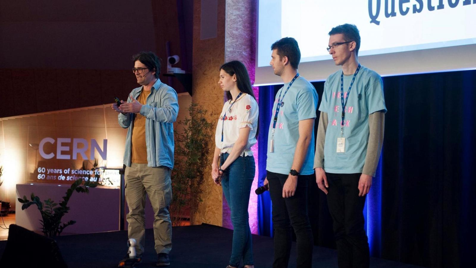
To solve this challenge and a series of others CERN organised a Hackathon for students and young professionals in April 2018 targeted to the medical field. RadiaBeam set the challenge of reimagining X-ray conversion and the selected team named HADDPHI (for Hardware Development Debrecen PHysics Institute) was given access to relevant CERN technologies.
Why is a hackathon organised by CERN focusing on MedTech? Early activities at CERN relating to medical applications date back to the 1970s. In light of the significant growth in these activities, in 2017, CERN published a formal medical applications strategy. The Medtech:Hack was then initiated to explore new ways of developing viable applications in the field.
So coming back to the RadiaBeam challenge: how to make X-ray conversion more efficient, reducing the size and cost of medical accelerators used for radiation therapy?
Before joining the Medtech:Hack, the HADDPHI team saw a presentation about Crystal Clear collaboration and were very impressed how High Energy Physics (HEP) can support medical applications. Though their exposure to the accelerators’ community was limited, they seized the opportunity to be exposed to more experts through the hack process.
From the CERN technologies, they selected the GEANT4 based on their past experience starting with simulations seemed a good idea. Furthermore, GEANT4 can be used to simulate one setup and its reliability has been proven in the field of particle physics. Finding the optimal configuration amongst millions needs the development of a framework that can manage this search. This could be performed thanks to the flexibility of the GEANT4 software.
So what is the solution the HADDPHI came up with?
Using GEANT to find the best parameters for an existing or paperboard X-ray devices for radiation therapy. It is key to find the optimal configuration amongst 100 to 200 parameters to render the X-ray spectrum as sharp as possible for X-ray therapy. They also investigated how to use the Timepix detector to monitor the beam created and therefore identify the corresponding delivered treatment to the patients.
When asked about their experience during the Hackathon, Berta Korcsmaros one of the team members stated: “We are electric engineers and physicists, we've never thought about business plan. Talking about how to bring a solution to the market was very new and very exciting to us. The Hackathon support team helped us a lot with making a good presentation. As well although we knew what CERN was doing, it was such a great experience to see how the Research and Development is done in real life.”
Indeed Timepix and GEANT4 experts explained to the team how to optimise the parameters to improve the Timepix detector and to use GEANT4 for the dedicated simulation to be performed. Also the Challenge Owner, Salime Boucher the CEO of RadiaBeam helped the team with benchmarking their proposed solution against state-of-the-art devices and this is him who introduced the team to the GEANT4 simulations. Moreover, for him, “The HADDPHI team came up with an interesting idea that I had not thought of, which was the use of the Timepix detector for realtime diagnosis of the beam energy. Currently, there is no direct way of measuring electron beam energy in medical linear accelerators. Instead we use indirect methods, such as the depth-dose profile of the resulting X-rays in water.”
With regards to the next steps, the team still has simulation work to do to find the optimal configuration for the accelerator generating the X-ray beam. In parallel, there will be the need to validate the simulation through detection measurements of the beam.
As Salime outlined: “At RadiaBeam we are in constant production of linear accelerators for medical and industrial applications. It would be quite easy for us to try out new X-ray converter geometries on one of our existing accelerators, so an experiment could be accomplished in just a few months. However, we probably would want to iterate between experiment and simulations a few times. ”
An interesting challenge ahead that will look familiar to any entrepreneurs trying to bring innovation out there!
Article originally published in Accelerating News.