Medipix in Medical Imaging
How does Medipix apply to medical imaging?
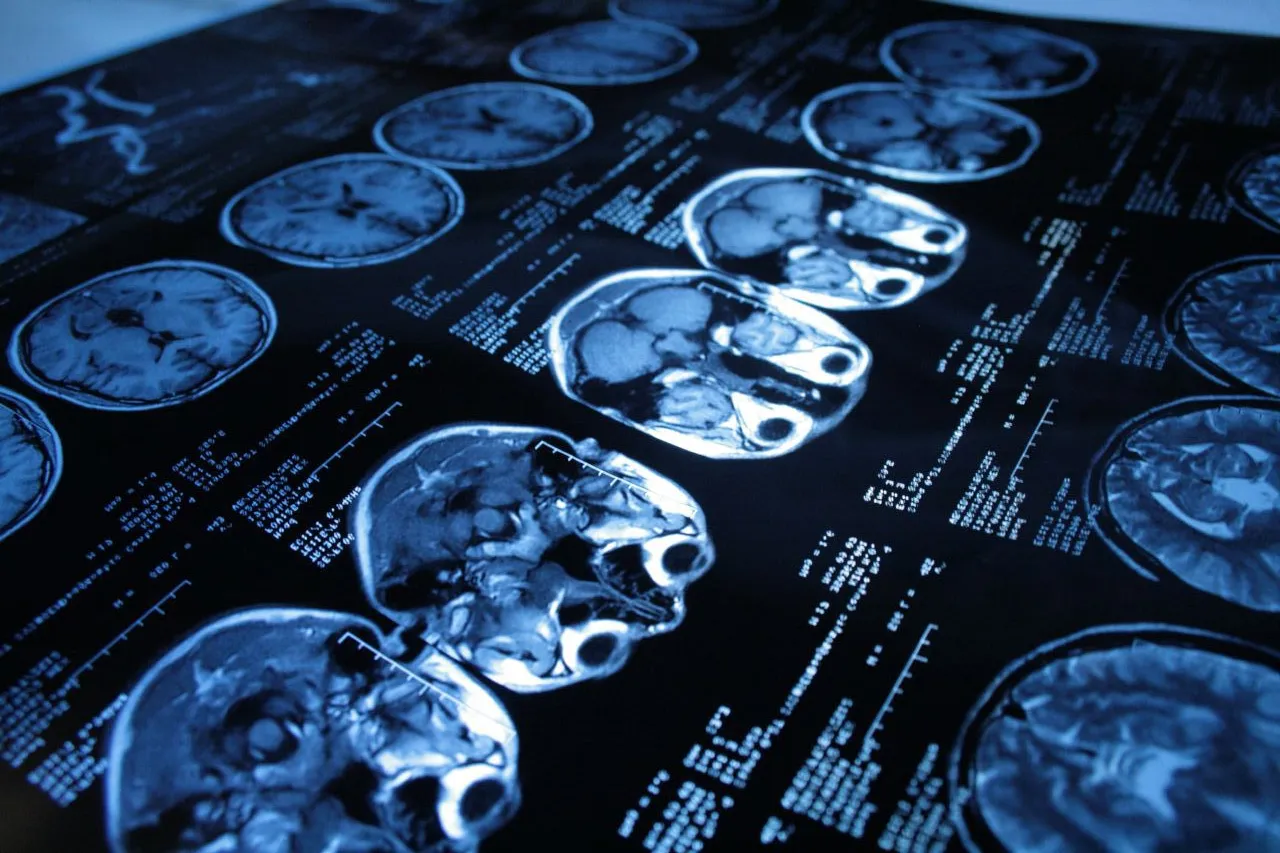
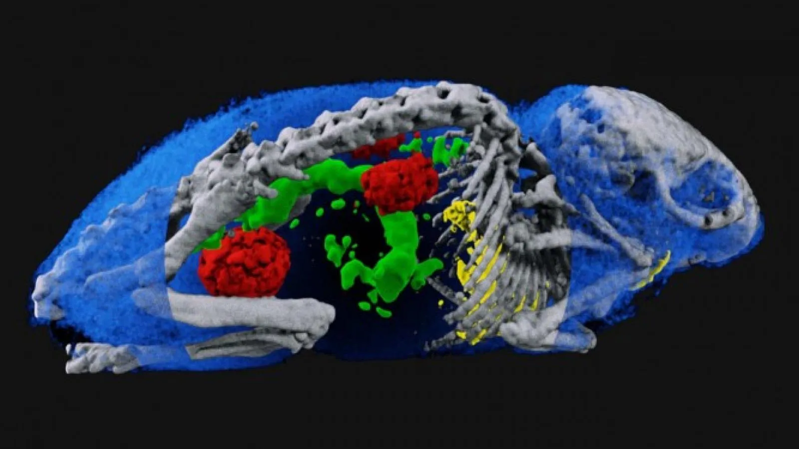
Radiography and computed tomography (CT) use X-ray photons to study the human body. The Medipix chips that implement on-pixel single photon counting provide many advantages for use in these fields. The technology has been applied in X-ray CT, in prototype systems for digital mammography, in CT imagers for mammography and for beta- and gamma-autoradiography of biological samples. Moreover, with the Medipix3 chip, the images are no longer black and white – they have colours to inducate different energy levels of the photons. The colour X-ray imaging technique produces clearer and more accurate pictures that should help doctors give their patients more accurate diagnoses.
Interested in getting a license in the area of medical imaging? Learn how here.
Projects in Medical Imaging:
Timepix Detector for Imaging in Ion Beam Radiotherapy
Ion beam radiotherapy enables better targeting radiation, sparing the surrounding tissue.
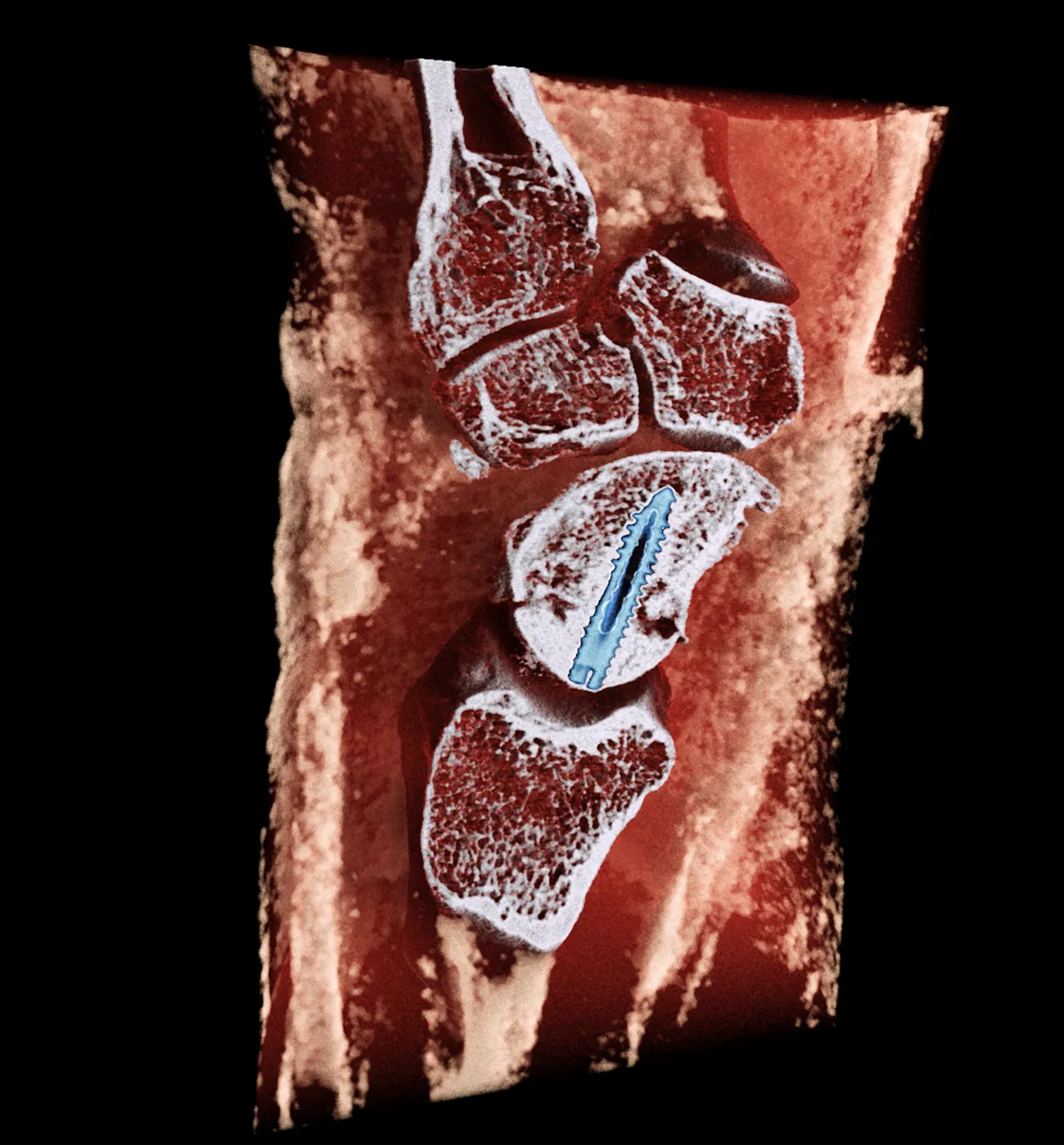
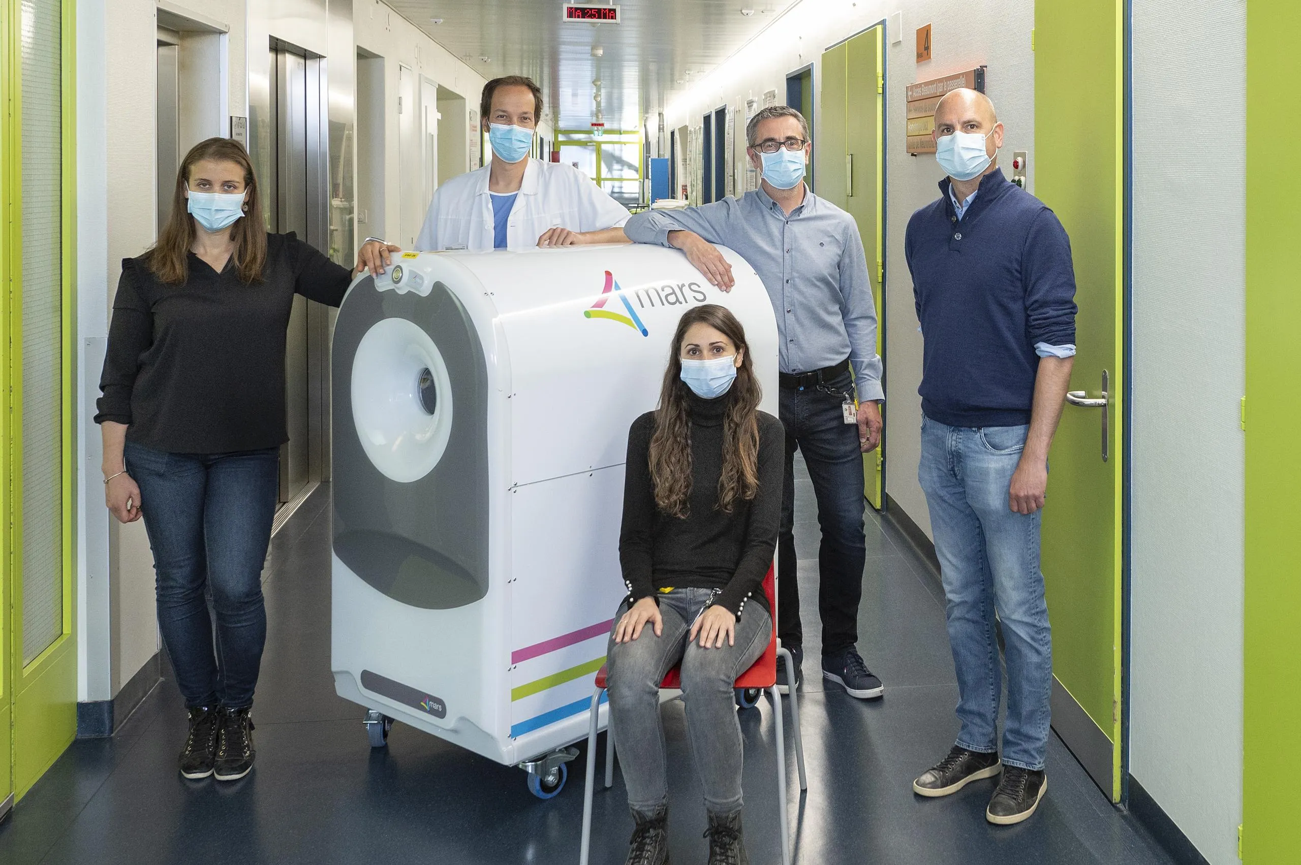
MARS Bioimaging partners with the Hospital for Special Surgery (HSS)
The partnership will assess particular aspects of the MARS 5×120 Extremity scanner.
Spectroscopic X-ray imaging now certified for medical use
The CERN Workshops on Medical Applications of Spectroscopic X-ray Detectors have been instrumental in advancing spectroscopic X-ray imaging and bringing it from the lab to the clinic
CERN’s impact on medical technology
Frontier instruments like the LHC and its detectors not only push back the boundaries of our knowledge, but also catalyse innovative technology for medical applications, writes Manuela Cirilli.
First European hospital receives 3D colour X-ray scanner using CERN technology
MARS Bioimaging’s 3D colour X-ray scanner has arrived in Europe to undertake clinical trials that will lead to its medical use.
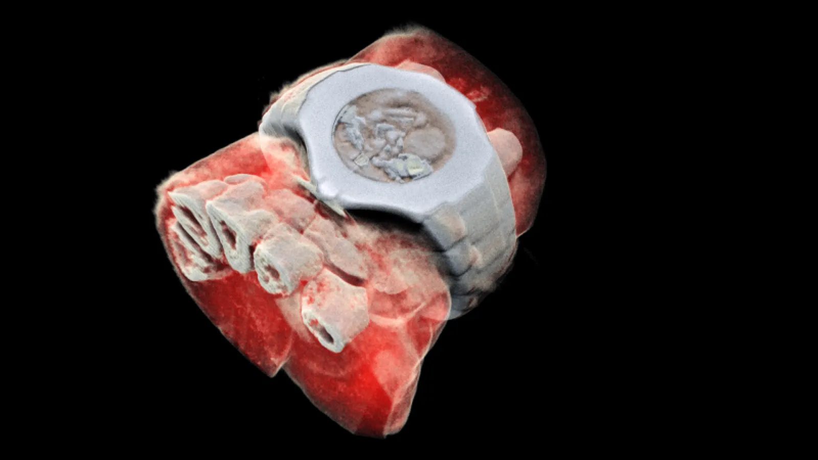
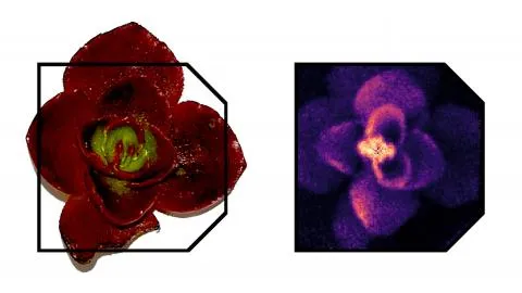
First 3D colour X-ray of a human using CERN technology
First human scanned with next–generation 3D colour scanner using CERN technology
Spectral imaging: from CERN to medical technologies
Ever since Röntgen discovered x-rays in 1895, physics and medicine have gone hand in hand, and advances made to particle detectors at CERN and elsewhere have continuously fuelled new developments in medical imaging.