MARS Bioimaging’s 3D colour X-ray scanner has arrived in Europe to undertake clinical trials that will lead to its medical use.
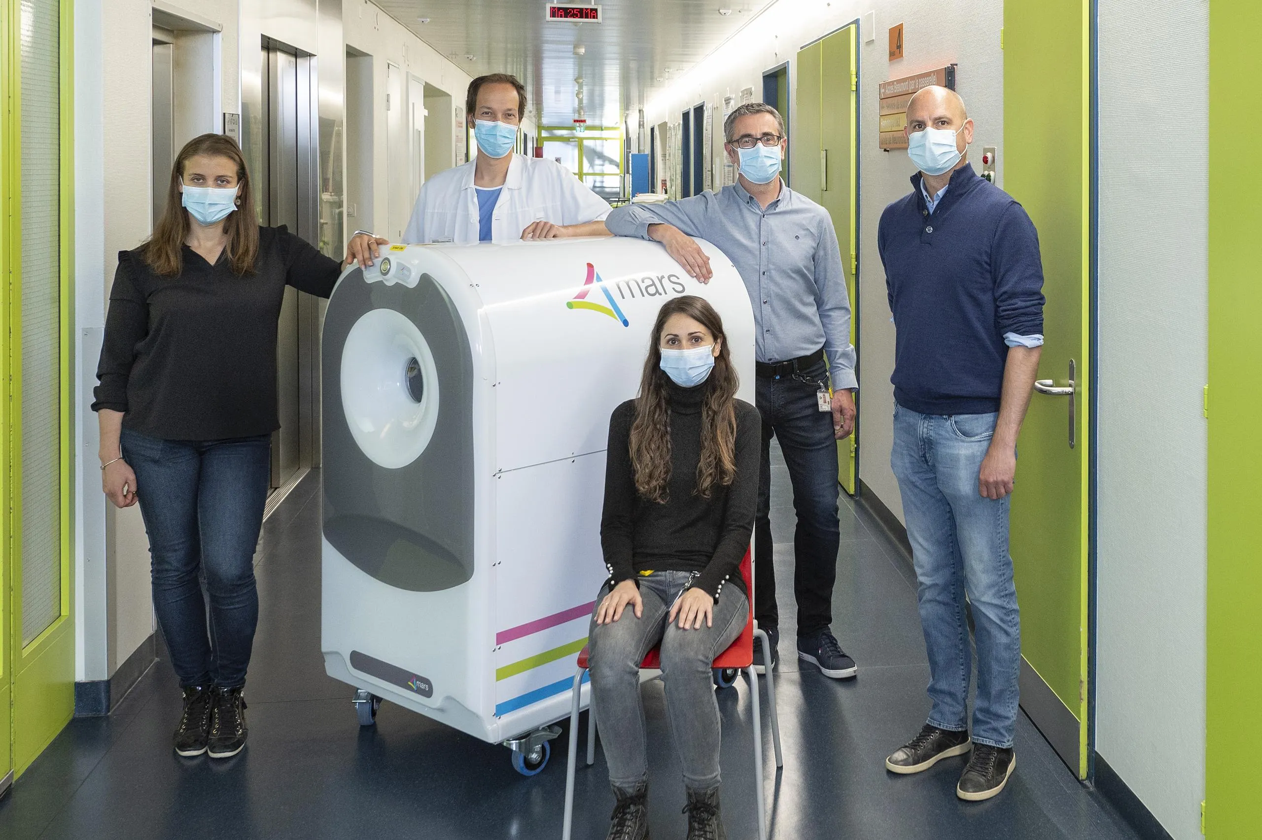
After years of development in New Zealand, MARS Bioimaging’s 3D colour X-ray scanner has arrived in Europe, at Lausanne University Hospital (CHUV) in Switzerland. This event marks the first step towards the European part of the international clinical trials undertaken by MARS Bioimaging, a New Zealand company, to obtain the certifications allowing its medical use.
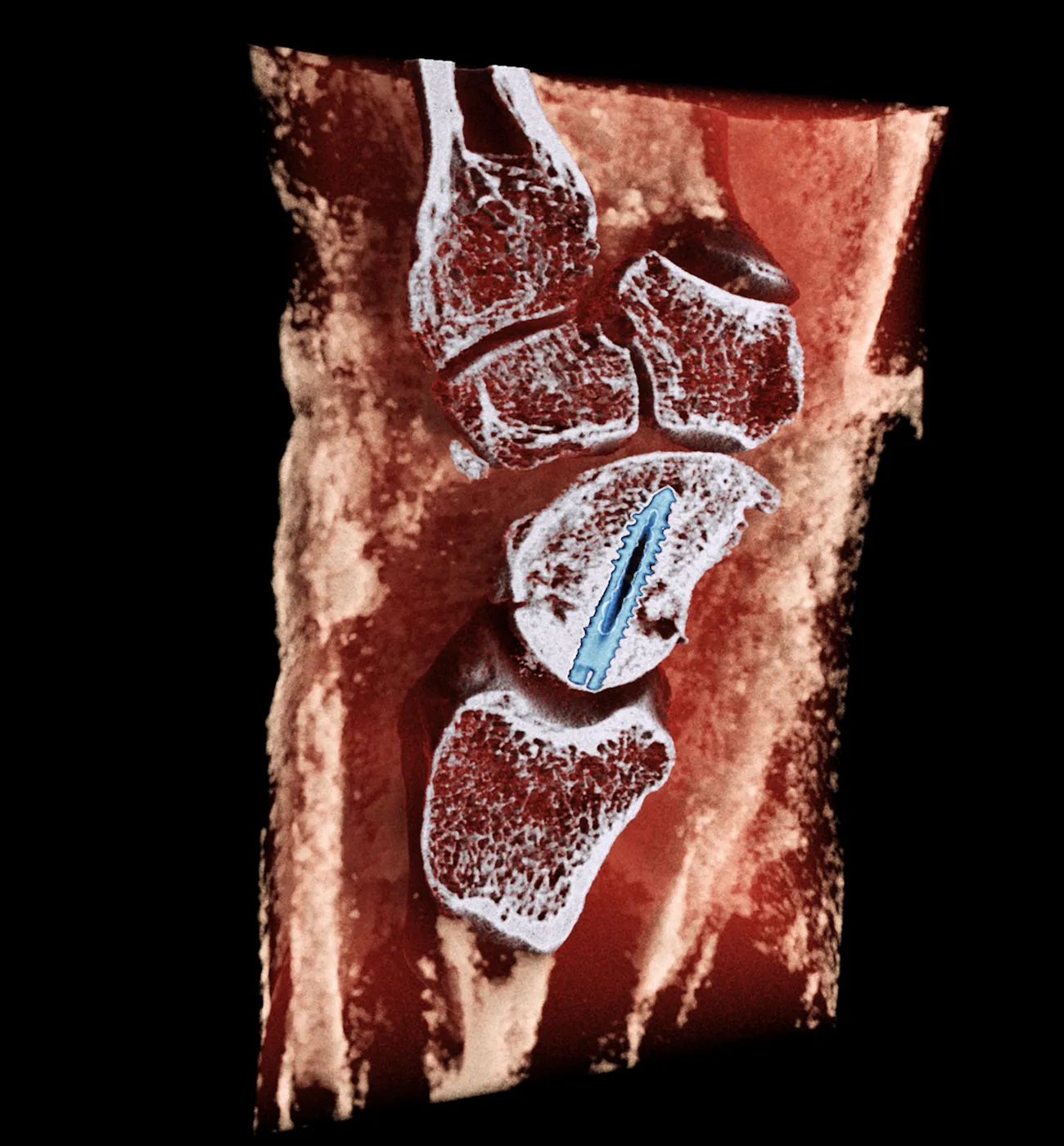
With the potential to monitor bone healing following a fracture, MARS Bioimaging’s scanner allows high resolution imaging around metal implants and can distinguish many different types of tissue without any use of contrast agents. Compared to existing technologies, images of such precision would enable significant progress to be made in diagnosing hand and wrist fractures and monitoring the healing process.
The entire team of radiologists and medical physicists at CHUV is eager to start the clinical use of the scanner. “The MARS scanner will allow us to improve our understanding of arthritis: how it develops and how to diagnose it. It should also help us develop the targeted therapies we are currently lacking for calcium crystal deposition diseases” explains Dr. Fabio Becce, Associate Physician and Senior Lecturer at Lausanne University Hospital (CHUV). “In practical terms, the MARS scanner gives us a unique opportunity to better identify and quantify urate and calcium crystal deposits in and around joints”.
“Reaching this incredible milestone is really exciting for us and our collaborators” says Professor Anthony Butler, President of MARS Bioimaging.
Trial of this technology in a Swiss hospital clearly demonstrates the pathway from experiments performed in a physics research laboratory to making a difference to patient health care
Professor Anthony Butler, President of MARS Bioimaging.
Since 2008, CERN and the New Zealand company have teamed up to develop a 3D colour X-ray scanner based on the Medipix3 technology, developed by the Medipix3 collaboration hosted by CERN. Inspired by particle physics detectors, Medipix3 and Timepix3 chips are now used for medical applications, in space, and for art authentication.
“In June 2021, CERN and MARS Bioimaging extended their current contract by five years, thus supporting MARS Bioimaging on their way to the US Food and Drug Administration and EU approval.”, explains Aurélie Pezous, from CERN’s Knowledge Transfer group. “The partnership between CERN, the Medipix3 Collaboration, and MARS Bioimaging shows how teaming up with health professionals is critical to enhance medical innovation.”
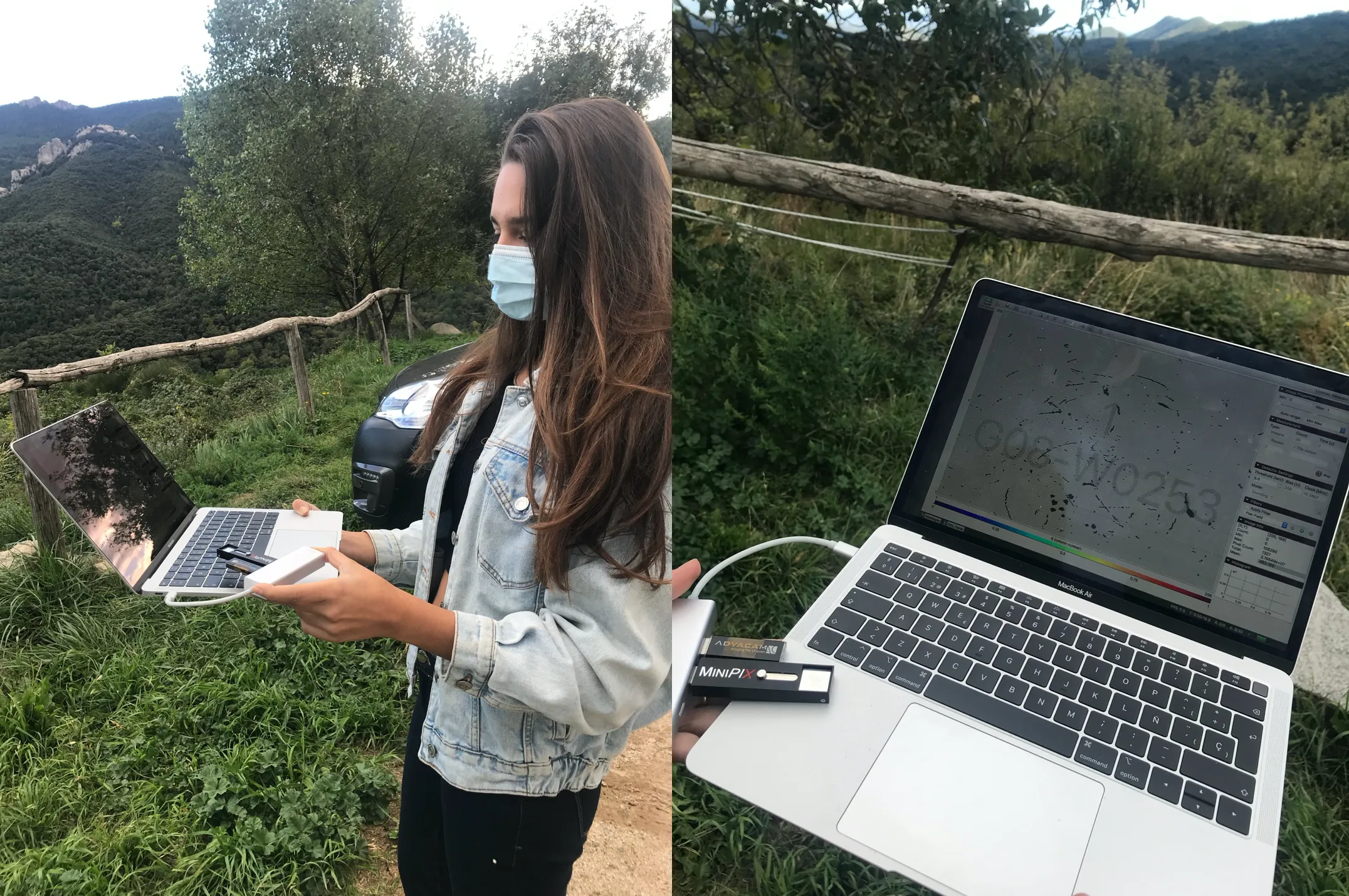
Beyond knowledge transfer, this cooperation highlights the potential of CERN Alumni. Various CERN Alumni have been involved in ensuring the scanner’s radiological safety. Amongst them is Lucia Gallego Manzano, former CERN fellow in radioprotection, now with the Institut de radiophysique (IRA) of CHUV.
Also On Medipix3
-
MARS Bioimaging partners with the Hospital for Special Surgery (HSS)
The partnership will assess particular aspects of the MARS 5×120 Extremity scanner. Read the original press release on the HSS website or our partner MBI’s website. Hospital for Special Surgery (HSS), the world’s leading academic medical center specialized in musculoskeletal health, and New Zealand-based medical device and technology company MARS Bioimaging Limited (MARS) today announced…
-
Source: CERN (CDS) Spectroscopic X-ray imaging now certified for medical use
The CERN Workshops on Medical Applications of Spectroscopic X-ray Detectors have been instrumental in advancing spectroscopic X-ray imaging and bringing it from the lab to the clinic BROKEN IMAGE NOT MOVED (drupal url : https://medipix.web.cern.ch//news/news/medipix3/spectroscopic-x-ray-imaging-now-certified-medical-use) The sixth Workshop on Medical Applications of Spectroscopic X-ray Detectors is currently taking place at CERN. Launched in 2011 and…
-
CERN’s impact on medical technology
Frontier instruments like the LHC and its detectors not only push back the boundaries of our knowledge, but also catalyse innovative technology for medical applications, writes Manuela Cirilli. This article was originally published in the July/August edition of CERN Courier magazine. Today, the tools of experimental particle physics are ubiquitous in hospitals and biomedical research.…
-
First European hospital receives 3D colour X-ray scanner using CERN technology
MARS Bioimaging’s 3D colour X-ray scanner has arrived in Europe to undertake clinical trials that will lead to its medical use. After years of development in New Zealand, MARS Bioimaging’s 3D colour X-ray scanner has arrived in Europe, at Lausanne University Hospital (CHUV) in Switzerland. This event marks the first step towards the European part…
-

Specifications
-
Features & Applications
Features & Applications Features: Applications:
-
Timepix-based detectors bring particle physics in the classroom
The ADMIRA project uses Timepix-based detectors to help students experiment with particle physics and contributes to transforming STEM education. Combustion, diffraction, magnetism… Many natural phenomena can be observed, experimented with and studied at school. Some have traditionally been difficult to demonstrate and have had to rely on theory only. Particle physics is one such subject…
