First human scanned with next–generation 3D colour scanner using CERN technology
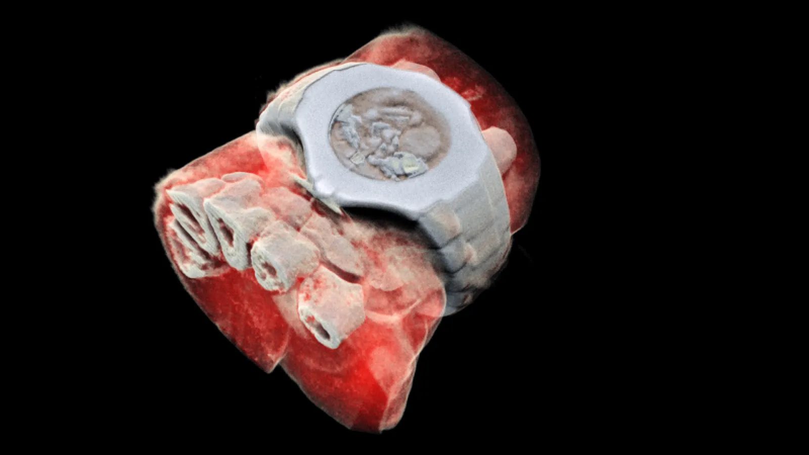
What if, instead of a black and white X-ray picture, a doctor of a cancer patient had access to colour images identifying the tissues being scanned? This colour X-ray imaging technique could produce clearer and more accurate pictures and help doctors give their patients more accurate diagnoses.
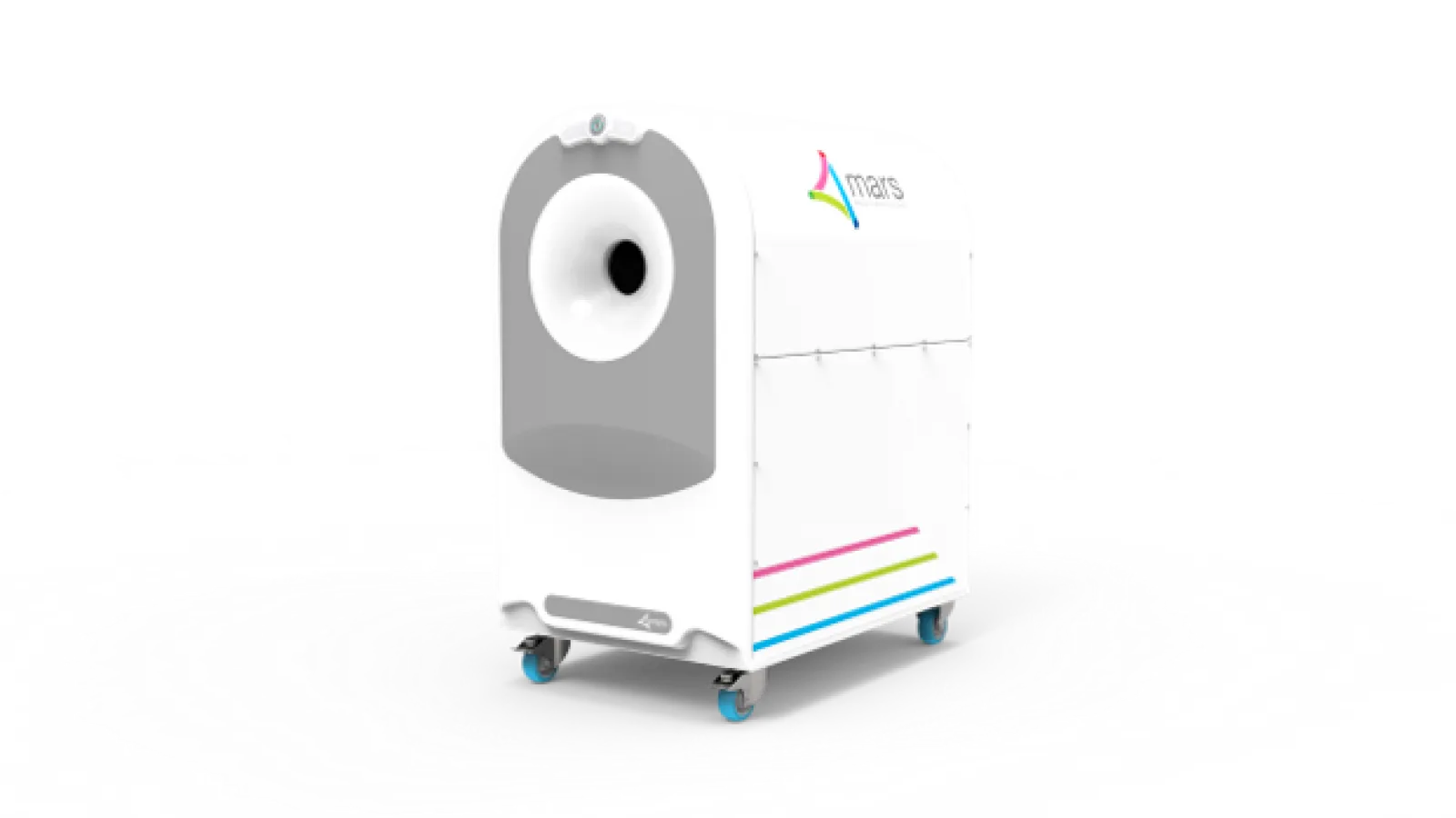
This is now a reality, thanks to a New-Zealand company that scanned, for the first time, a human body using a breakthrough colour medical scanner based on the Medipix3 technology developed at CERN. Father and son scientists Professors Phil and Anthony Butler from Canterbury and Otago Universities spent a decade building and refining their product.
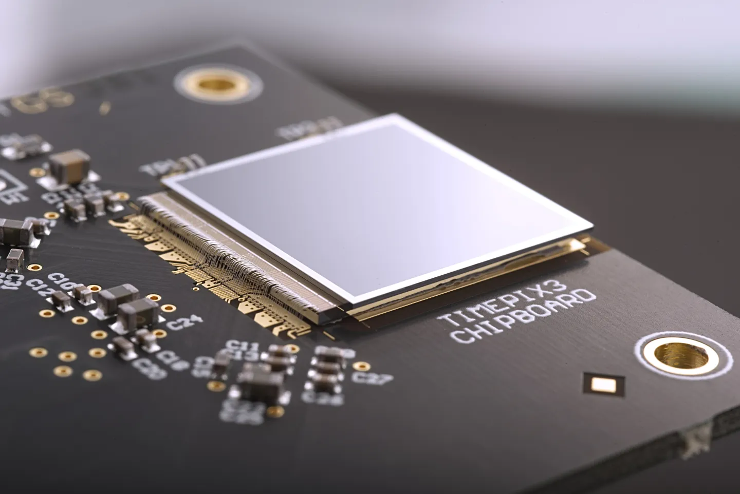
Medipix is a family of read-out chips for particle imaging and detection. The original concept of Medipix is that it works like a camera, detecting and counting each individual particle hitting the pixels when its electronic shutter is open. This enables high-resolution, high-contrast, very reliable images, making it unique for imaging applications in particular in the medical field.
Hybrid pixel-detector technology was initially developed to address the needs of particle tracking at the Large Hadron Collider, and successive generations of Medipix chips have demonstrated over 20 years the great potential of the technology outside of high-energy physics.
MARS Bioimaging Ltd, which is commercialising the 3D scanner, is linked to the University of Otago and Canterbury. The latter together with more than 20 research institutes forms the third generation of Medipix collaboration. The Medipix3 chip is the most advanced chip available today and Professor Phil Butler recognises that “this technology sets the machine apart diagnostically because its small pixels and accurate energy resolution mean that this new imaging tool is able to get images that no other imaging tool can achieve.”
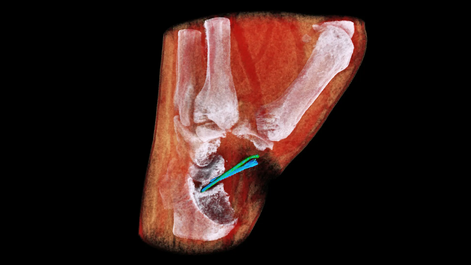
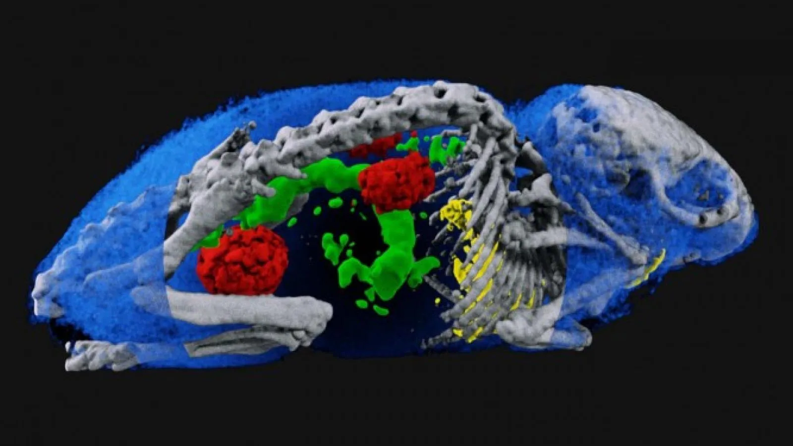
MARS’ solution couples the spectroscopic information generated by the Medipix3 enabled detector with powerful algorithms to generate 3D images. The colours represent different energy levels of the X-ray photons as recorded by the detector hence identifying different components of body parts such as fat, water, calcium, and disease markers.
So far, researchers have been using a small version of the MARS scanner to study cancer, bone and joint health, and vascular diseases that cause heart attacks and strokes. “In all of these studies, promising early results suggest that when spectral imaging is routinely used in clinics it will enable more accurate diagnosis and personalisation of treatment,” Professor Anthony Butler says.
CERN’s Knowledge Transfer group has a long-standing expertise in transferring CERN technologies, in particular for medical applications. In the case of the 3D scanner, a license agreement has been established between CERN, on behalf of Medipix3 collaboration and MARS Bioimaging Ltd. As Aurélie Pezous, CERN Knowledge Transfer Officer states, “It is always satisfying to see our work leveraging benefits for patients around the world. Real-life applications such as this one fuels our efforts to reach even further.”
In the coming months, orthopaedic and rheumatology patients in New Zealand will be scanned by the revolutionary MARS scanner in a clinical trial that is a world first, paving the way to a potentially routine use of this new generation equipment.
Also On Medipix
-

Specifications
-
Features and Applications
Features: Applications:
-
New 3D colour X-rays made possible with CERN technology
Stunning new images pave the way to large-scale human trials, two years on from the first ever 3D colour human X-ray using CERN Medipix3 technology Two years after the first ever 3D colour X-ray of a living human, MARS Bioimaging have released stunning new images made using a world-first compact scanner, based on Medipix3 technology…
-
MARS Bioimaging is seeking partners to better enable drug development and therapy monitoring for COVID-19
The current standard for definitive diagnosis of COVID-19 is the real-time reverse transcriptase-polymerase chain reaction (RT-PCR). Background The current standard for definitive diagnosis of COVID-19 is the real-time reverse transcriptase-polymerase chain reaction (RT-PCR). While this is a highly sensitive and specific test for COVID-19, it does not measure the severity nor progress of the associated…
-
Medipix: Two decades of turning technology into applications
The story of how detector components ended up in medical imaging, in art restoration and even in space. How could microchips developed for detectors at the Large Hadron Collider (LHC) be used beyond high-energy physics? This was a question that led to the Medipix and Timepix families of pixel-sensor chips. Researchers saw many possible applications…
-
First 3D colour X-ray of a human using CERN technology
First human scanned with next–generation 3D colour scanner using CERN technology What if, instead of a black and white X-ray picture, a doctor of a cancer patient had access to colour images identifying the tissues being scanned? This colour X-ray imaging technique could produce clearer and more accurate pictures and help doctors give their patients…
-
How to spot a perfect fake: the world’s top art forgery detective
BBC on Fake Art – InsightART in the report! BBC on Fake Art – InsightART in the report! In: BBC News, May 12th Also On Medipix
-
Spectral imaging: from CERN to medical technologies
Ever since Röntgen discovered x-rays in 1895, physics and medicine have gone hand in hand, and advances made to particle detectors at CERN and elsewhere have continuously fuelled new developments in medical imaging. Ever since Röntgen discovered x-rays in 1895, physics and medicine have gone hand in hand, and advances made to particle detectors at…
-
Particle detectors for the classroom
Physics teacher Becky Parker of the Physics Simon Langton Grammar School in the UK keeps particle detectors in her classroom. Physics teacher Becky Parker of the Physics Simon Langton Grammar School in the UK keeps particle detectors in her classroom. For years, her students have been using Medipix detector – silicon-pixel particle detectors that fit…
-
Medipix: From particles to patients
In the 1990s, researchers on the Large Hadron Collider were assembling the last pieces of detector electronics, designed to capture high-resolution images of particle collisions quickly and with no background noise. In the 1990s, researchers on the Large Hadron Collider were assembling the last pieces of detector electronics, designed to capture high-resolution images of particle…