The current standard for definitive diagnosis of COVID-19 is the real-time reverse transcriptase-polymerase chain reaction (RT-PCR).
Background
The current standard for definitive diagnosis of COVID-19 is the real-time reverse transcriptase-polymerase chain reaction (RT-PCR). While this is a highly sensitive and specific test for COVID-19, it does not measure the severity nor progress of the associated lung disease.
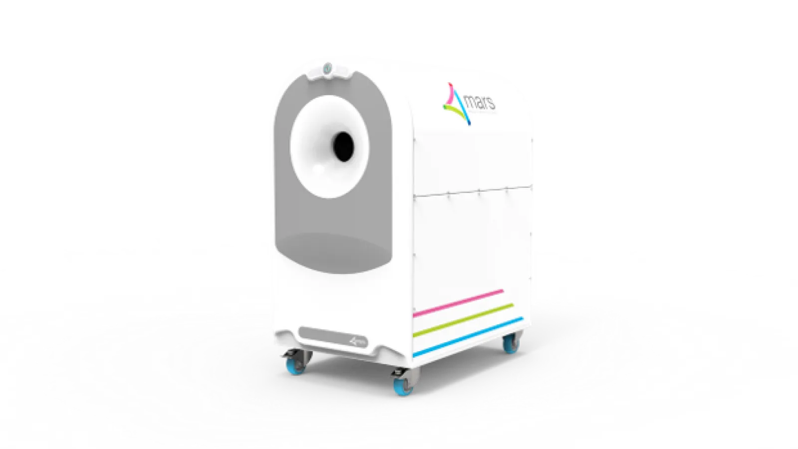
Significance of CERN Technology/MARS imaging could help COVID-19 treatment monitoring and drug development
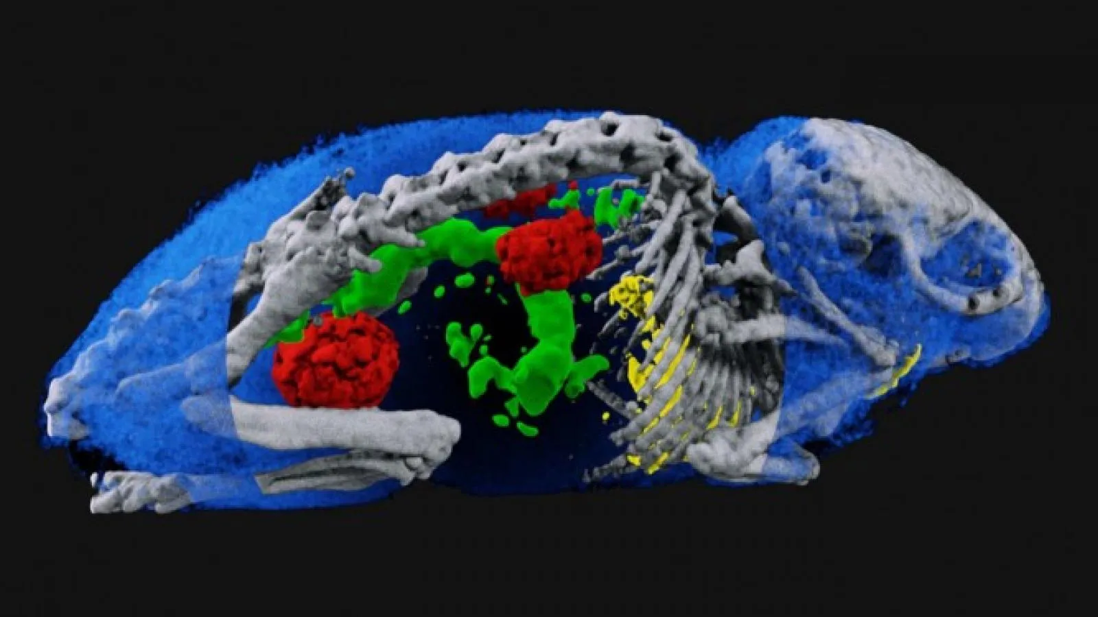
Mars technology utilises the varying absorption of X-rays across the diagnostic energy range. This enables MARS to generate functional imaging by simultaneously identifying and quantifying various components of tissues as well as exogenously administered pharmaceutical agents such as nanoparticles. Hence there is the possibility of having a tagging agent for lung injury and fibrosis that can be visualised. This would enable both highly specific and quantitative imaging for either Covid-19 or the lungs response to the virus. Developing a technique would enable better diagnosis, drug development, and monitoring of therapy. This applies to both animal trials for drug research and human clinical trials.
Proposal
The MARS team in collaboration with its various partners have demonstrated that spectral photon-counting for molecular imaging is useful for detecting lung TB using iodine and silver contrast agents, bone micro-fractures using hafnium particles, and HER-2 positive cell lines using gold nanoparticles in breast cancer research1, 2, 3. It is hoped that a similar quantitative marker can be found for assessing Covid-19 lung damage. They are seeking potential partners who have access to, or capable of, developing disease models like and/or Covid. Interested parties may contact Professor Anthony Butler at anthony.butler@marsbioimaging.com or www.marsbioimaging.com for further information.
Publications:
1. https://doi.org/10.1002/adfm.201904936
2. https://doi.org/10.1007/978-981-32-9816-3_17
3. https://www.ncbi.nlm.nih.gov/pmc/articles/PMC4958312/
Also On Medipix
-

Specifications
-
Features and Applications
Features: Applications:
-
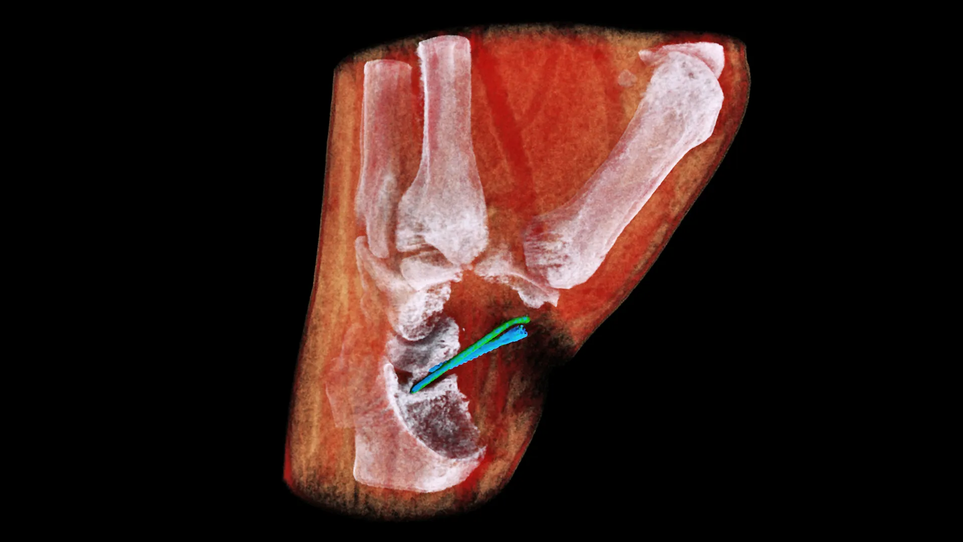
New 3D colour X-rays made possible with CERN technology
Stunning new images pave the way to large-scale human trials, two years on from the first ever 3D colour human X-ray using CERN Medipix3 technology Two years after the first ever 3D colour X-ray of a living human, MARS Bioimaging have released stunning new images made using a world-first compact scanner, based on Medipix3 technology…
-
MARS Bioimaging is seeking partners to better enable drug development and therapy monitoring for COVID-19
The current standard for definitive diagnosis of COVID-19 is the real-time reverse transcriptase-polymerase chain reaction (RT-PCR). Background The current standard for definitive diagnosis of COVID-19 is the real-time reverse transcriptase-polymerase chain reaction (RT-PCR). While this is a highly sensitive and specific test for COVID-19, it does not measure the severity nor progress of the associated…
-
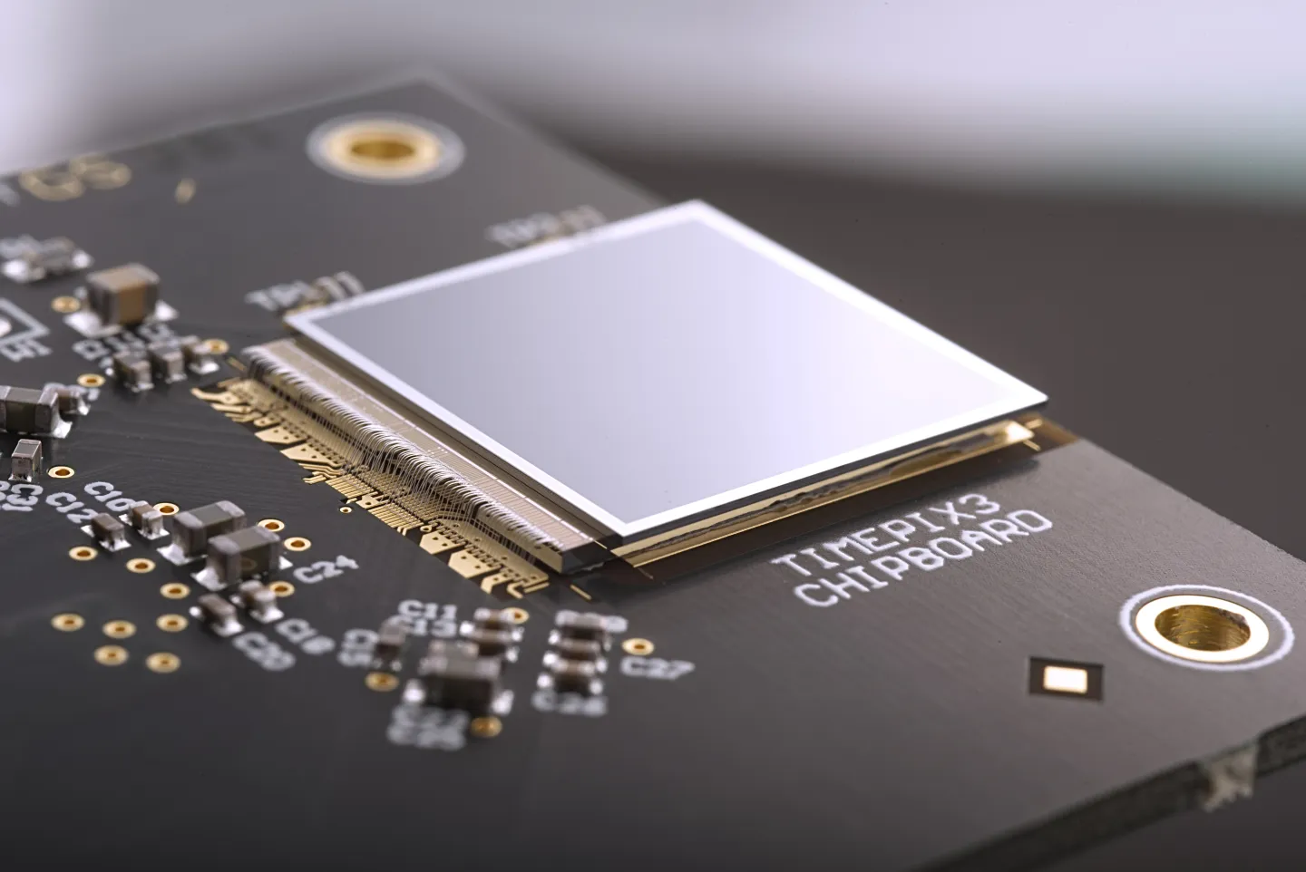
Medipix: Two decades of turning technology into applications
The story of how detector components ended up in medical imaging, in art restoration and even in space. How could microchips developed for detectors at the Large Hadron Collider (LHC) be used beyond high-energy physics? This was a question that led to the Medipix and Timepix families of pixel-sensor chips. Researchers saw many possible applications…
-
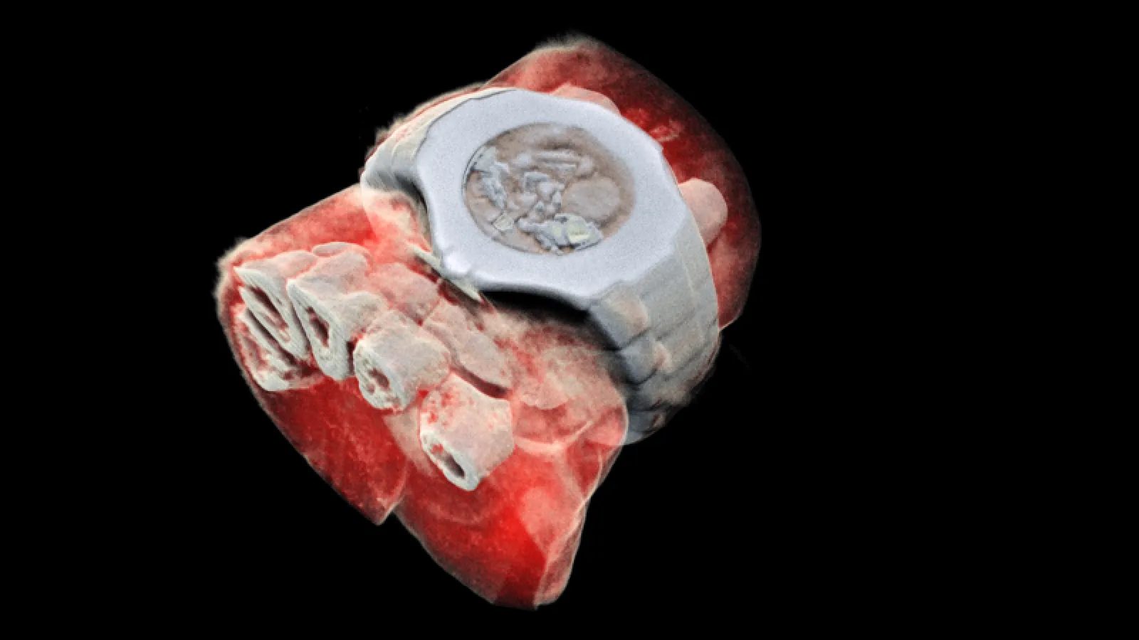
First 3D colour X-ray of a human using CERN technology
First human scanned with next–generation 3D colour scanner using CERN technology What if, instead of a black and white X-ray picture, a doctor of a cancer patient had access to colour images identifying the tissues being scanned? This colour X-ray imaging technique could produce clearer and more accurate pictures and help doctors give their patients…
-
How to spot a perfect fake: the world’s top art forgery detective
BBC on Fake Art – InsightART in the report! BBC on Fake Art – InsightART in the report! In: BBC News, May 12th Also On Medipix
-
Spectral imaging: from CERN to medical technologies
Ever since Röntgen discovered x-rays in 1895, physics and medicine have gone hand in hand, and advances made to particle detectors at CERN and elsewhere have continuously fuelled new developments in medical imaging. Ever since Röntgen discovered x-rays in 1895, physics and medicine have gone hand in hand, and advances made to particle detectors at…
-
Particle detectors for the classroom
Physics teacher Becky Parker of the Physics Simon Langton Grammar School in the UK keeps particle detectors in her classroom. Physics teacher Becky Parker of the Physics Simon Langton Grammar School in the UK keeps particle detectors in her classroom. For years, her students have been using Medipix detector – silicon-pixel particle detectors that fit…
-
Medipix: From particles to patients
In the 1990s, researchers on the Large Hadron Collider were assembling the last pieces of detector electronics, designed to capture high-resolution images of particle collisions quickly and with no background noise. In the 1990s, researchers on the Large Hadron Collider were assembling the last pieces of detector electronics, designed to capture high-resolution images of particle…